

Abstract
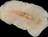
This study examined the ultrastructural characteristics of the egg of Coquillettidia venezuelensis (Theobald) (Diptera: Culicidae) with the aid of scanning electron microscopy. The eggs are elliptical and blackish, measuring on average 525.3 ± 12.8 μm in length and 94.9 ± 5.14 μm in width, with a length to width ratio of 5.54 ± 0.18. The anterior region of the egg is tubuliform, with a well-developed micropylar collar measuring approximately 2.54 ± 0.33 μm in thickness, the inner side of which is uniformly and deeply excavated, with a depression around the outer margin of the micropylar disc, which has a diameter of 29.4 ± 1.7 μm, an area of 234.4 μm2 ± 401.3 μm2 and a slightly elevated central region measuring 1.7 ± 0.5 μm in diameter that bears the central micropyle.
References
Alencar, J., Gil-Santana, H.R., de Mello, C.F., Marcondes, C.B. & Dos Santos-Mallet, J.R. (2019) Ultrastructure and morphometry of the egg of Coquillettidia albifera (Prado) with illustrations of male genitalia (Diptera: Culicidae). Zootaxa, 4565 (1), 145–150.
https://doi.org/10.11646/zootaxa.4565.1.13
Bates, M. & Roca-Garcia, M. (1945) Laboratory studies of the Saimiri-Haemagogus cycle of jungle yellow fever. American Journal of Tropical Medicine, 25, 203–216.
https://doi.org/10.4269/ajtmh.1945.s1-25.203
Belkin, J.N., Heinemann, S.J. & Page, W.A. (1970) The Culicidae of Jamaica (Mosquito studies. XXI). Contributions of the American Entomological Institute, 6 (1), 1–458.
Clark-Gil, S. & Darsie Jr., R.F. (1983) The mosquitoes of Guatemala. Mosquito Systematics, 13, 151–284.
Consoli, R.A.G.B. & De Oliveira, R.L. (1994) Principais mosquitos de importância sanitária no Brasil. Editora FIOCRUZ, Rio de Janeiro, 228 pp.
https://doi.org/10.7476/9788575412909
Cova García, P., Sutil, E. & Rausseo J.A. (1966). Mosquitos (Culicinos) de Venezuela. Vol. I & II. Ministerio de Sanidad y Asistencia Social, Caracas, 410 + 413 pp.
de Mello, C.F., dos Santos-Mallet, J.R., Morone, F., Guimarães, A.É., Marcondes, C.B. & Alencar, J. (2014) Ultrastructure of the egg of Coquillettidia juxtamansonia (Chagas, 1907) (Diptera: Culicidae). Journal of Vector Ecology, 39, 219–221.
https://doi.org/10.1111/j.1948-7134.2014.12090.x
dos Santos-Mallet, J.R., Gleiser, R.M., Alencar, J., Marques, W.D.A., Sarmento, J.S., Müller, G.A. & Marcondes, C.B. (2009) Scanning electron microscopy of the egg of Ochlerotatus albifasciatus (Diptera: Culicidae). Journal of Medical Entomology, 46, 980–985.
https://doi.org/10.1603/033.046.0502
Forattini, O.P. (2002) Culicidologia Médica. 2nd Edition. EDUSP, São Paulo, 864 pp.
Harbach, R.E. (2020a) Valid Species. Mosquito Taxonomic Inventory. Available from: http://mosquito-taxonomic-inventory.info/valid-species-list (accessed 10 September 2020)
Harbach, R.E. (2020b) Anatomical glossary. Mosquito Taxonomic Inventory. Available from: http://mosquito-taxonomic-inventory.info/node/11027 (accessed 10 September 2020)
Hervé, J.P., Dégallier, N., Travassos da Rosa, A.P.A., Pinheiro, F.P. & Sá Filho, G.C. (1986) Arboviroses - Aspectos ecológicos. In: Fsesp, I. (Ed.), Instituto Evandro Chagas—50 anos de contribuição às ciências biológicas e à medicina tropical. Vol. I. Fundação Serviços de Saúde Pública, Belém, pp. 409–437
Lane, J. (1953) s.n. In: Neotropical Culicidae. Vol. II. University of São Paulo, São Paulo, pp. 553–1112.
Lounibos, L.P., Duzak, D. & Linley, J.R. (1997) Comparative egg morphology of six species of the Albimanus Section of Anopheles (Nyssorhynchus) (Diptera: Culicidae). Journal of Medical Entomology, 34, 136–155.
https://doi.org/10.1093/jmedent/34.2.136
Mattingly, P.F. (1971) Mosquito eggs XVIII. Genus Mansonia (subgenera Rhynchotaenia Brèthes and Mansonia Blanchard) with a further note on genus Ficalbia Theobald. Mosquito Systematics, 4, 45–49.
Motta, M.A., Lourenço-de-Oliveira, R. & Sallum, M.A.M. (2007) Phylogeny of genus Wyeomyia (Diptera: Culicidae) inferred from morphological and allozyme data. Canadian Entomologist, 139, 591–627.
https://doi.org/10.4039/n06-088
Reinert, J.F. (2009) List of abbreviations for currently valid generic-level taxa in family Culicidae (Diptera). Journal of the European Mosquito Control Association, 27, 68–76.
Reinert, J.F. (2010) Species of mosquitoes (Diptera: Culicidae) with published illustrations and/or descriptions of eggs − Summary. European Mosquito Bulletin, 28, 182–186.
Reinert, J.F., Harbach, R.E. & Kitching, I.J. (2004) Phylogeny and classification of Aedini (Diptera: Culicidae), based on morphological characters of all life stages. Zoological Journal of the Linnean Society, 142, 289–368.
https://doi.org/10.1111/j.1096-3642.2004.00144.x
Westphal-Ferreira, B., Vieira, T.B., da Silva, A.M. & Navarro-Silva, M.A. (2018) Scanning electron microscopy of the eggs of Coquillettidia shannoni (Lane & Antunes, 1937) and Phoniomyia quasilongirostris (Theobald, 1907) (Diptera: Culicidae). Journal of Vector Ecology, 43, 193–197.
https://doi.org/10.1111/jvec.12300
White, G.B. & Faust, C. (2014) Appendix 4. Medical acarology and entomology. In: Farrar, J., Hotez, P., Junghanss, T., Kang, G., Lalloo, D. & White, N.J. (Eds.), Manson’s Tropical Infectious Diseases. 23rd Edition. Elsevier Saunders Ltd., Philadelphia, pp. 1258–1272.