

Abstract
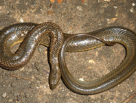
Based on two male and two female individuals, we describe a new genus and species of mud snake, Myanophis thanlyinensis gen. nov., sp. nov., from the vicinity of the campus of East Yangon University, Yangon, Thanlyin, Myanmar. This species differs from every other homalopsid species by the following combination of characters: (1) dorsal scales smooth, row formula 21–21–19 or 21–21–17; (2) tail short, ratio tail length/SVL 0.185–0.204 in males, 0.160–0.167 in females; (3) nasal scales separated; (4) 125–126 ventral scales in males, 120–122 in females; (5) 38–39 subcaudal scales in males, 32–34 in females; and (6) hemipenis bilobed. Its matrilineal genealogy (based on analyses of 16S and cytochrome b sequences), associates Myanophis thanlyinensis gen. nov., sp. nov. most closely with species of the genera Myrrophis and Gyiophis. The new taxon differs from the species of Myrrophis and Gyiophis by having a bilobed hemipenis (vs. unilobed). Myanophis thanlyinensis gen. nov., sp. nov. differs further from the species of Myrrophis by having 125–126 ventral scales in males and 120–122 in females (vs. 137–162 and 137–164, respectively), and 38–39 subcaudal scales in males and 32–34 in females (vs. 39–55 and 37–52, respectively). Myanophis thanlyinensis gen. nov., sp. nov. differs further from the species of Gyiophis by lacking dark blotches along flank (vs. present), and by having 21 dorsal scales rows at midbody (vs. 25). We provide an identification key to the homalopsid species known to occur in Myanmar. As a novelty to the classic holotype description and characterization, the individual has been genome sequenced by Illumina short-read technology and its genome has been assembled into a draft nuclear genome and a complete, annotated mitochondrial genome. This innovative approach comprehensively and permanently characterizes the genomic variation of the holotype.
References
Marçais, G., Kingsford, C. (2011) A fast, lock-free approach for efficient parallel counting of occurrences of k-mers. Bioinformatics, 27 (6), 764–770.
https://doi.org/10.1093/bioinformatics/btr011
Vurture, G.W., Sedlazeck, F.J., Nattestad, M., Underwood, C.J., Fang, H., Gurtowski, J. & Schatz, M.C. (2017) GenomeScope: fast reference-free genome profiling from short reads. Bioinformatics, 33 (14), 2202–2204.
https://doi.org/10.1093/bioinformatics/btx153
Bolger, A.M., Lohse, M. & Usadel, B. (2014) Trimmomatic: a flexible trimmer for Illumina sequence data. Bioinformatics, 30 (15), 2114–2120.
https://doi.org/10.1093/bioinformatics/btu170
Wood, D.E., Lu, J. & Langmead, B. (2019) Improved metagenomic analysis with Kraken 2. Genome biology, 20 (1), 257.
https://doi.org/10.1186/s13059-019-1891-0
Bankevich, A., Nurk, S., Antipov, D., Gurevich, A.A., Dvorkin, M., Kulikov, A.S., Lesin, V.M., Nikolenko, S.I., Son, P., Prjibelski, A.D., Pyshkin, A.V., Sirotkin, A.V., Vyahhi, N., Tesler, G., Alekseyev, M.A. & Pevzner, P.A. (2012) SPAdes: a new genome assembly algorithm and its applications to single-cell sequencing. Journal of computational biology, 19 (5), 455–477.
https://doi.org/10.1089/cmb.2012.0021
Li H (2013) Aligning sequence reads, clone sequences and assembly contigs with BWA-MEM. arXiv:1303.3997v2
Li, H., Handsaker, B., Wysoker, A., Fennell, T., Ruan, J., Homer, N., Marth, G., Abecasis, G. & Durbin, R. (2009) The sequence alignment/map format and SAMtools. Bioinformatics, 25 (16), 2078–2079.
https://doi.org/10.1093/bioinformatics/btp352
Okonechnikov, K., Conesa, A. & García-Alcalde, F. (2016) Qualimap 2: advanced multi-sample quality control for high-throughput sequencing data. Bioinformatics, 32 (2), 292–294.
https://doi.org/10.1093/bioinformatics/btv566
Quinlan, A.R. & Hall, I.M. (2010) BEDTools: a flexible suite of utilities for comparing genomic features. Bioinformatics, 26 (6), 841–842.
https://doi.org/10.1093/bioinformatics/btq033
R Core Team (2020) R: A language and environment for statistical computing. R Foundation for Statistical Computing, Vienna, Austria. [https://www.R-project.org/]
Meng, G., Li, Y., Yang, C. & Liu, S. (2018) MitoZ: A toolkit for mitochondrial genome assembly, annotation and visualization. bioRxiv, 489955.
https://doi.org/10.1101/489955
Dierckxsens, N., Mardulyn, P. & Smits, G. (2017) NOVOPlasty: de novo assembly of organelle genomes from whole genome data. Nucleic acids research, 45 (4), e18–e18.
https://doi.org/10.1093/nar/gkw955
Sievers, F., Wilm, A., Dineen, D., Gibson, T.J., Karplus, K., Li, W., Lopez, R., McWilliam, H., Remmert, M., Söding, J., Thompson, J.D. & Higgins, D.G. (2011) Fast, scalable generation of high-quality protein multiple sequence alignments using Clustal Omega. Molecular systems biology, 7(1), 539.
https://doi.org/10.1038/msb.2011.75
Tillich, M., Lehwark, P., Pellizzer, T., Ulbricht-Jones, E. S., Fischer, A., Bock, R. & Greiner, S. (2017) GeSeq–versatile and accurate annotation of organelle genomes. Nucleic acids research, 45 (W1), W6–W11.
https://doi.org/10.1093/nar/gkx391
Donath, A., Jühling, F., Al-Arab, M., Bernhart, S.H., Reinhardt, F., Stadler, P.F., Middendorf, M. & Bernt, M. (2019) Improved annotation of protein-coding genes boundaries in metazoan mitochondrial genomes. Nucleic acids research, 47 (20), 10543–10552.
https://doi.org/10.1093/nar/gkz833
Chan, P.P., & Lowe, T.M. (2019) tRNAscan-SE: searching for tRNA genes in genomic sequences. In: Gene Prediction. Humana, New York, NY. pp. 1–14.
https://doi.org/10.1007/978-1-4939-9173-0_1
Chan, P.P., Lin, B.Y., Mak, A.J. & Lowe, T.M. (2019) tRNAscan-SE 2.0: improved detection and functional classification of transfer RNA genes. bioRxiv, 614032.
https://doi.org/10.1101/614032
Laetsch, D.R. & Blaxter, M.L. (2017) BlobTools: Interrogation of genome assemblies. F1000Research, 6( 1287), 1287.
https://doi.org/10.12688/f1000research.12232.1
Simão, F.A., Waterhouse, R.M., Ioannidis, P., Kriventseva, E.V., Zdobnov, E.M. (2015) BUSCO: assessing genome assembly and annotation completeness with single-copy orthologs. Bioinformatics, 31 (19), 3210–3212.
https://doi.org/10.1093/bioinformatics/btv351